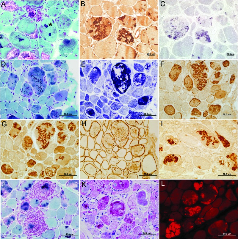
Figure 2. Histologic and immunocytochemical findings.
(A, D, J) Pleomorphic granular or hyaline material and cytoplasmic bodies in the abnormal fibers in trichrome-stained sections. Note a nucleus surrounded by dark blue granular material (asterisk, A) and inflammatory exudate surrounding and replacing abnormal fibers (J). (E) The hyaline masses are devoid of NADH dehydrogenase activity. (K) Periodic acid-Schiff–positive material appears near inclusions. (L) Fibers harbor intensely congophilic inclusions. Nonconsecutive sections in the same series show abnormal fiber regions that immunoreact strongly for FHL1 (B) and part of the same regions react for menadione–nitro blue tetrazolium without substrate (C). Note abnormal accumulation of αB-crystallin (F), myotilin (G), dystrophin (H), and ubiquitin (I) in the structurally abnormal fibers. Samples were from the right soleus muscle of patient 4 (A) and from the right triceps muscle of patient 2 (B–L). (A, D–L) Scale bar = 50 μm. (B, C) Scale bar = 20 μm.

