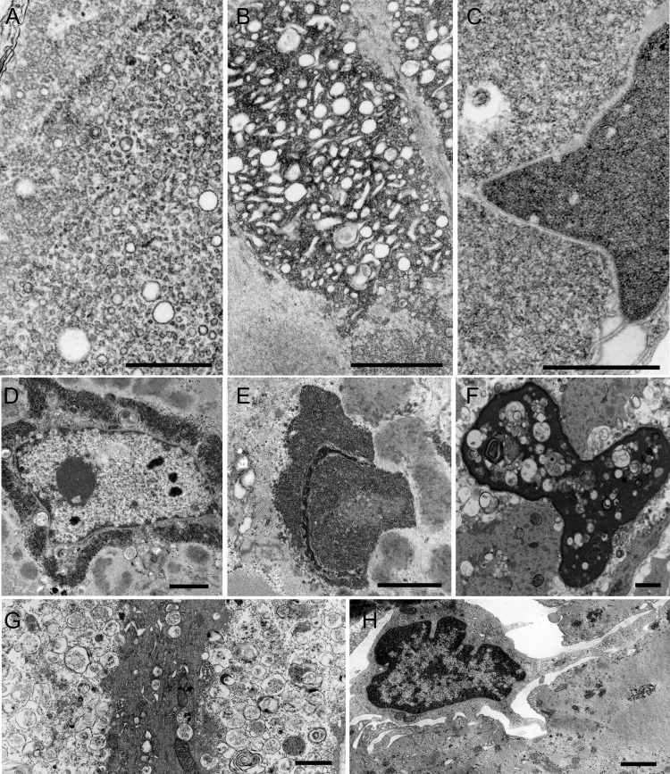
Figure 4. Electron microscopy findings.
(A) Aggregate of rough endoplasmic reticulum profiles. (B) Clusters of dilated sarcotubular profiles intermingled with tubulofilamentous material. (C–F) Nuclear alterations. (C, E) The nuclei are filled with the tubulofilamentous material. (D) This material encases the nucleus. A matrix of irregularly oriented fine filaments intermingled with ribosomes is also seen (C). (F) A highly degenerate nucleus is filled with multiple small myeloid structures and debris. (G) A highly degenerate fiber containing remnants of myofibrils surrounded by degenerating organelles. (H) An abnormal fiber is invaded by a mononuclear cell. (A) Scale bar = 0.5 μm. (B–H) Scale bar = 1 μm in (B–H).

