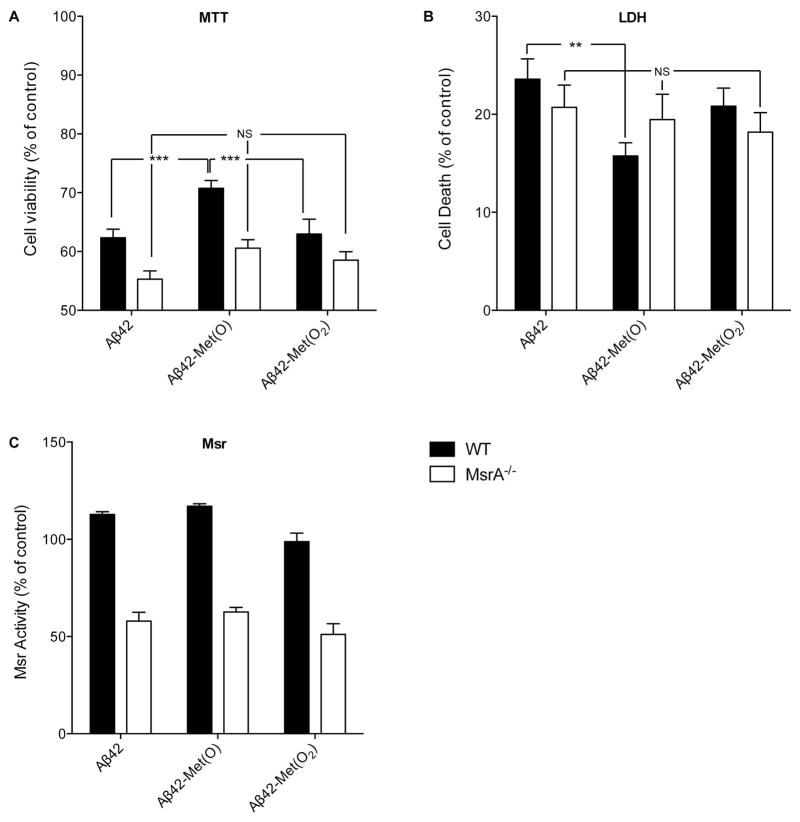
Figure 3. Response of MSRA−/− and WT mouse primary cortical neurons to native and oxidized Aβ42.
Primary MsrA−/− or WT mouse cortical neurons were treated with 10 μM of each Aβ analogues for 48h. A) Assessment of cell viability using the MTT assay. B) Assessment of cell death using the LDH assay. C) Measurement of specific Msr activity by HPLC using dabsyl-Met(O) as substrate. The results were normalized to untreated WT cells (150 pmol dabsyl-Met/min/mg protein defined as 100% specific Msr activity). The data are an average of 5–10 independent experiments. **p < 0.01, ***p < 0.001, NS – non-significant.

