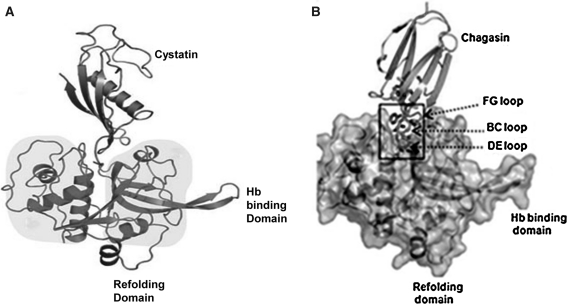
Fig. 8.
a 3D structure of falcipain 2–cystatin complex. Cystatin is colored in orange, and falcipain-2 protease is colored in green. Refolding domain and hemoglobin binding domain highlighted in pink and salmon, respectively (Figure has been taken from Wang et al. 2006). b Structure of falcipain 2–chagasin complex: overall structure of chagasin with falcipain-2, chagasin in red and falcipain 2 in golden color (Figure has been taken from Wang et al. 2007). (Color figure online)

