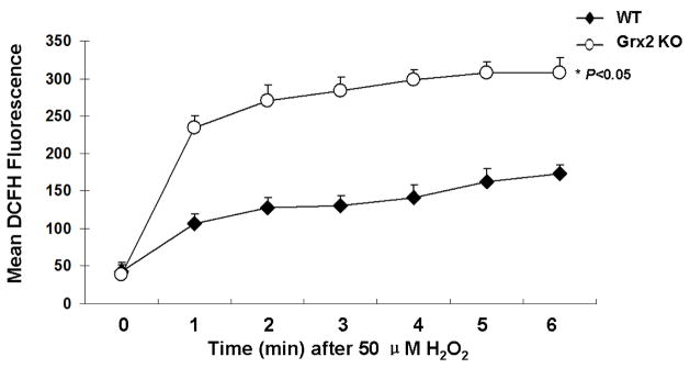
Figure 3. H2O2 detoxification in wild type and Grx2 knockout lens epithelail cells.
Wild type (WT) and Grx2 knockout (Grx2 KO) primary mouse LECs were plated in 96-well plate (all normalized to 10,000 cells/well) and treated with PBS containing 50 μM DCFH-DA. After a baseline was acquired, 50 μM H2O2 was added to the cells and DCF fluorescence levels were determined at given time points. Error bars indicate S.D., n=6, *P<0.05 vs. WT.

