Figure 1.

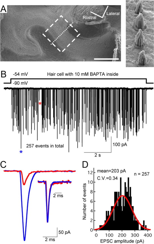
Hair cells from the bullfrog amphibian papilla: a step depolarization evokes multivesicular EPSCs even when the hair cell is dialyzed with 10 mm BAPTA. A, Scanning electron micrograph of the amphibian papilla surface, showing the 400–500 Hz region where electrophysiological recordings and morphological reconstructions were performed (dashed box). The higher magnification image (right) shows hair bundles from hair cells along the dotted line. Scale bar: 200 μm. B, Paired recordings of a single 10 mm BAPTA dialyzed hair cell and its connected afferent fiber show that a step depolarization of the hair cell from −90 to −54 mV was sufficient to evoke large-amplitude EPSCs that occurred stochastically during the depolarizing pulse. C, A large-amplitude EPSC (blue star) and a small-amplitude EPSC (red star) from B are superimposed and shown at high temporal resolution. The two EPSCs have similar waveforms after normalization to the same peak value (inset). D, Amplitude distribution of all EPSCs shown in B (bin size, 5 pA). The distribution can be fit with a Gaussian function (red) and has an average amplitude of 203 pA (n = 257 EPSCs), corresponding to a quantal content of approximately four to five quanta (or released vesicles).
