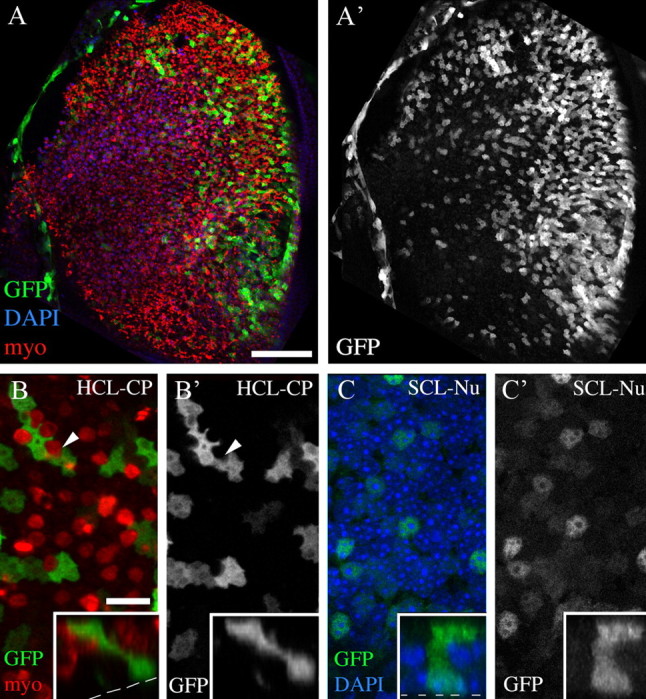
Figure 2.

Adenovirus transduces supporting cells but not hair cells. All colored images show labeling for GFP (green), myosin VIIa (myo; red), and/or DAPI (blue) from utricles that were explanted, treated overnight with Ad5-CMV-GFP in media lacking neomycin, cultured for an additional day, and fixed. Individual grayscale panels show labels as indicated. A, A′, Brightest point projections of z-series through the SE at low magnification. A′, GFP labeling in same field as A. B, B′, A higher-magnification slice through the hair cell layer at the level of cuticular plate (HCL-CP), near the cell's top at the organ's lumen. B, GFP and myo labeling; B′, GFP only. Arrowheads in B and B′ point to a myosin VIIa+ top of a hair cell lacking GFP signal. C, C′, A slice through the supporting cell layer at the level of the nucleus (SCL-Nu) in the same field as B and B′. C, GFP and DAPI labeling; C′, GFP only. Insets in B–C′ show vertical slices (z/y) through two GFP+ cells to illustrate their elongated shapes that are typical of supporting cells. Scale bars: A, A′ (in A), 100 μm; B–C′ (in B), 6 μm.
