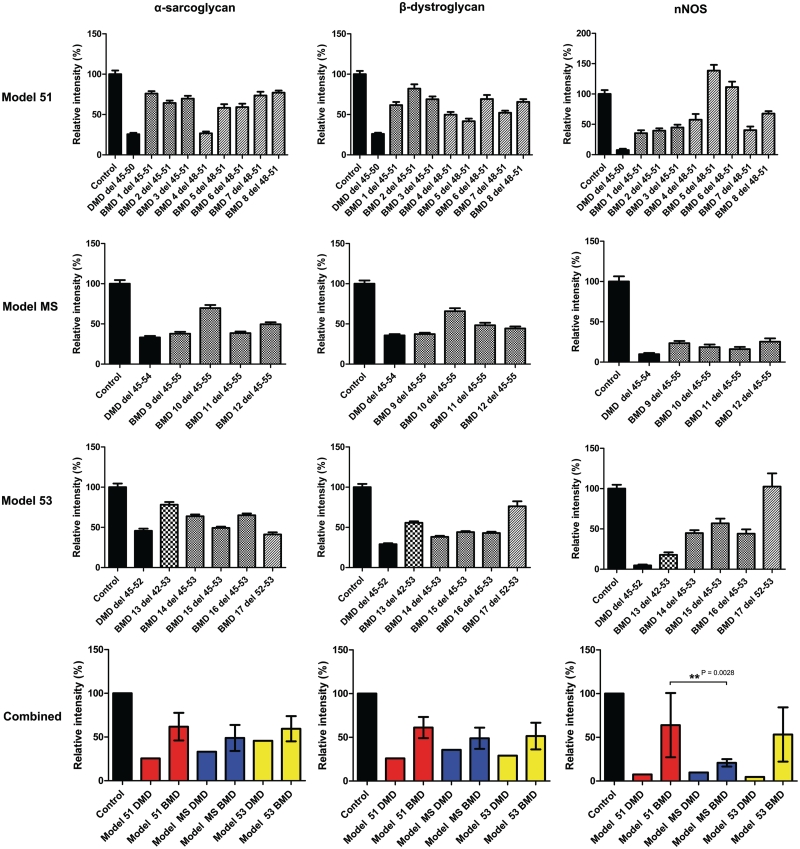
Figure 4.
Comparative immunohistochemical analysis of dystrophin-associated protein expression in 17 patients with Becker muscular dystrophy (BMD) with in-frame deletions. Control, Duchenne muscular dystrophy and Becker muscular dystrophy transverse muscle sections were immunolabelled with antibodies against α-sarcoglycan, β-dystroglycan, neuronal nitric oxide synthase (nNOS) and β-spectrin. Expression was quantified relative to control muscle in 40 muscle fibres and normalized to β-spectrin expression. Values represent means ± SEM except for graphs on the bottom where values represent the mean expression level for each group ± SD of the difference between sample means. In the bottom two graphs patients with Becker muscular dystrophy are grouped according to corresponding exon skipping models for Duchenne muscular dystrophy: exon 51 skipping (model 51, red bars), multi-exon skipping (model MS, blue bars) and exon 53 skipping (model 53, yellow bars).

