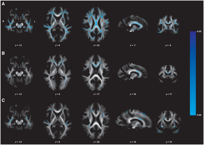
Figure 2.
Neuroimaging results of the brain (DTI, group comparison of fractional anisotropy values). Displayed results of tract-based spatial statistics analyses of fractional anisotropy values (group comparison) are based on a corrected threshold of Pfamily wise error < 0.05. Mean tract-based spatial statistics tract skeleton is overlaid on the mean fractional anisotropy image (display threshold of 0.1). The coordinates refer to the MNI reference space. (A) Fractional anisotropy reduction in patients with myotonic dystrophy type 1 compared with healthy controls. (B) Fractional anisotropy reduction in patients with myotonic dystrophy type 2 compared with healthy controls. (C) Fractional anisotropy reduction in patients with myotonic dystrophy type 1 compared with patients with myotonic dystrophy type 2.

