Figure 3.

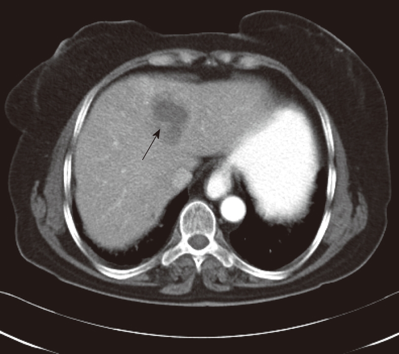
A 70-year-old female patient presented with right upper abdominal pain lasting 16 wk. Abdominal computerized tomographic examination showed low density masses with hazy margins located to medial segment of the left lobe (arrow).

A 70-year-old female patient presented with right upper abdominal pain lasting 16 wk. Abdominal computerized tomographic examination showed low density masses with hazy margins located to medial segment of the left lobe (arrow).