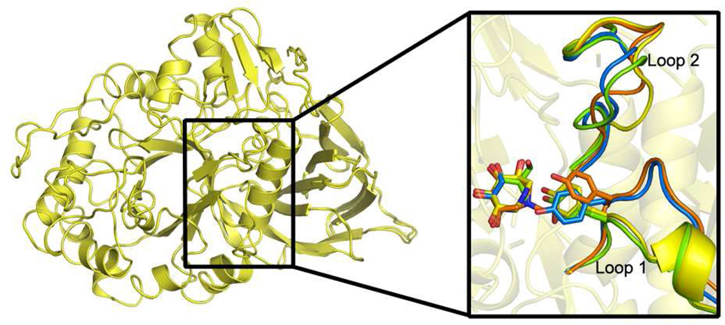
Fig. 4.
Superposition of 1 and 2 bound GCase structures and comparison of loops adjacent to the active site (inset). After binding, Loop 1 adopts either a helical turn as seen for compound 1 (inset, yellow) and IFG (inset, green), or an extended loop conformation seen in the compound 2 (inset, orange) and glycerol (inset, blue) bound structures. Changes in Loop 2 are due to crystal packing (see text).

