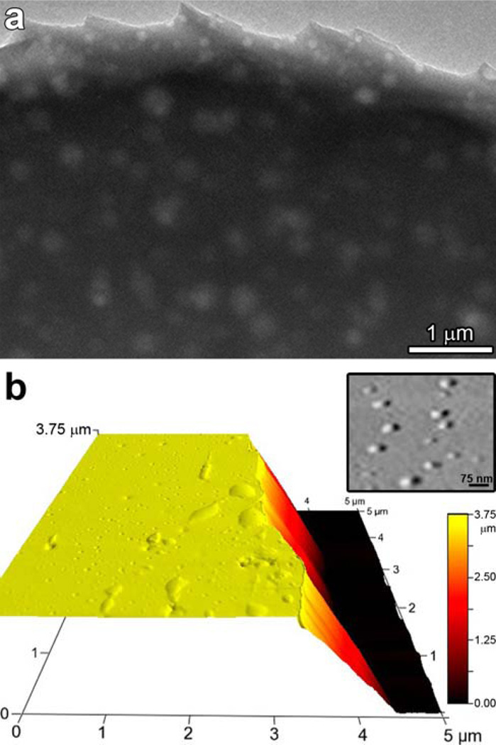
Figure 10.
(a) Transmission electron micrograph of printed porous pigment which was then removed from the polymer substrate and placed on a TEM grid. The 50 to 200 nm features show the porosity created in these ormosil xerogel spots, which are ~4 µm thick and 1 mm in diameter. (b) An AFM micrograph in perspective showing the height of the porous pigment at the spot center compared to the base height of the PET as revealed by a scrape from a blade. Inset shows an enlargement of the surface pores (< 100 nm) (from ref. 69).

