
Figure 5.
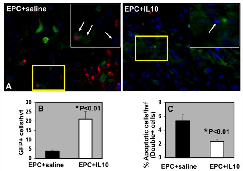
A. EPC retention and survival in the myocardium at 3 days post-MI in IL-10/saline treated mice. TUNEL staining for detecting apoptosis (Red) of EPC (GFP-positive, green fluorescence) and DAPI (blue) for nuclear staining. Inset is higher magnification of the yellow-boxed area. Arrows indicate GFP+TUNEL+ cells. B. Quantification of GFP+ (EPC) cells at 3 days post-MI. C. Quantitative analysis of GFP/TUNEL double-positive cells at 3 days post-MI. IL-10 increased GFP+ EPC retention and survival in the heart following transplantation, *P<0.01 vs EPC+saline group. hvf, high-power visual field.