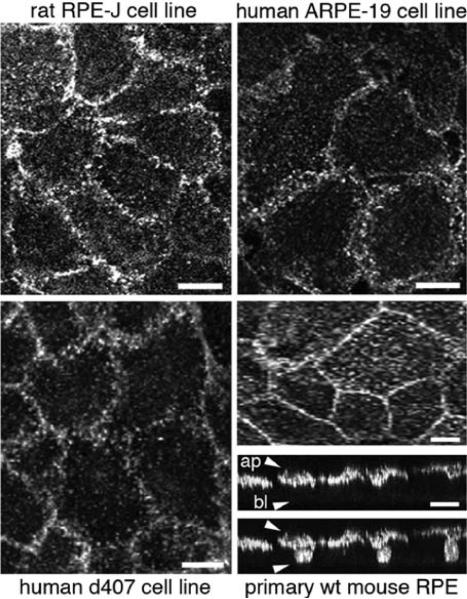
Fig.15.1.
Apical polarity of αvβ5 integrin receptors in RPE cells in culture. Rat RPE-J, human ARPE-19 and human d407 RPE cell lines, as indicated, were grown to confluence on glass coverslips and labeled live on ice with αvβ5 surface dimer-specific antibody P1F6. 3D projections representing the upper 2 μm of apical aspects of cells are shown. Wild-type 129 strain mouse RPE was isolated in patches from 10-day-old mouse eyes, cultured for 4 days before fixation and labeling with antibody recognizing the β5 integrin cytoplasmic domain. Images were acquired by laser scanning confocal microscopy. Representative whole cell maximal projections of the same field are shown in x–y plane and in x–z plane. x–z projection is shown with (upper panel) and without (lower panel) nuclei counterstaining. Approximate locations of apical (ap) and basolateral (bl) surfaces of cells are indicated by arrowheads in the upper panel. Scale bar is 10 μm for cell lines and 20 μm for primary RPE

