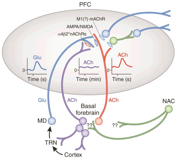
Figure 2.
Circuitry model describing the main components of the prefrontal cortex (PFC) circuitry mediating signal detection and processing mode shifts. The model combines evidence with parsimonious assumptions required to explain electrochemical and attentional performance data (see main text for details). The glutamatergic (GLU) inputs to the PFC, originating from the mediodorsal thalamic nucleus (MD) “import” a preattentionally processed representation of the signal into the PFC (see text for definition). MD neurons are part of a network that includes the thalamic reticular nucleus (TRN) and its topographic afferents from sensory cortical regions. The cue-evoked glutamatergic transient (see insert) generates a cholinergic transient (see insert), via stimulation of ionotropic presynaptic glutamate receptors (Parikh, et al., 2010; Parikh, et al., 2008). This cholinergic transient mediates the actual detection process or, depending on the task, a processing mode shift that fosters detection (see main text). Prefrontal output neuron activity is stimulated by ACh primarily via muscarinic (m)AChRs, thereby organizing the behavioral responses that indicate successful detection.
The terminals of the MD inputs to the PFC are equipped with α4β2* nAChRs. Cholinergic stimulation of these receptors is thought to vary over minutes, refecting a tonic component of cholinergic neurotransmission (see elevated release illustrated by the insert). nAChR agonists enhance detection performance primarily by positively modulating GLU release from these terminals, thereby augmenting the amplitudes of the cholinergic transients (Howe, et al., 2010; Parikh, et al., 2010). This model therefore proposes two separate roles for cholinergic inputs, mediated via separate populations of cholinergic neurons. A rather tonically active cholinergic input modulates glutamate release from MD neurons which, in turn, target the terminals of a separate group of cholinergic neurons, generating the transients that enhance attentional orienting and cue detection. Reproduced, with permission of Nature Publishing Group, from Hasselmo and Sarter (2011; p. 58).

