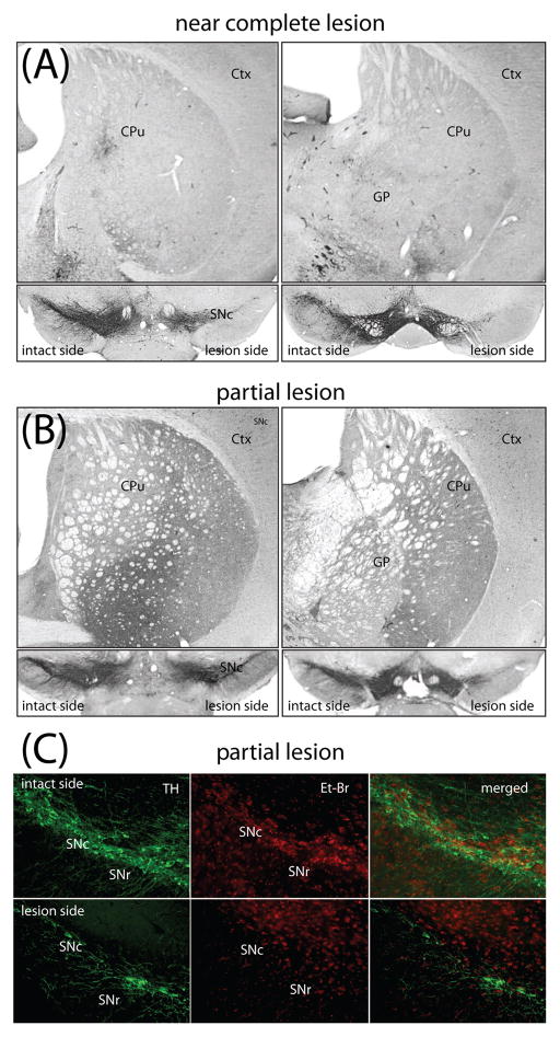
Fig 1.
Near complete lesion and partial nigrostriatal lesion induced with different 6-OHDA doses. (A) Unilateral intrastriatal injection of 3.5 μg 6-OHDA caused a near complete elimination of TH-immunoreactive fibers throughout the rostro-caudal axis of lateral striatum (A) and a near complete loss of SNc DA cells (B). SNc is shown in coronal sections at different levels of mouse midbrain in rostro-caudal order. (C) Fluorescence microphotographs of TH positive neurons counterstained with ethidium bromide in coronal sections through the midbrain of mice injected with 2.5 μg 6-OHDA. On the side contralateral to the lesion (intact side) the SNc contained a normal number of large neurons with dendrites positive for TH fluorescence. Ethidium bromide counterstaining overlaped with TH positive neurons. Ipsilateral to the injection site (lesion side) there were few viable neurons labeled with TH and ethidium bromide fluorescence. Ctx - cortex, CPu-caudate putamen (striatum), GP-globus pallidus, VTA-ventral tegmental area, SNc – substantia nigra pars compacta, SNr - substantia nigra pars reticulata, TH – tyrosine hydroxylase, Et-Br - ethidium bromide.

