
Figure 1. Low concentrations of DHPs suppress dendritic Ca2+ oscillations but do not slow pacemaking.
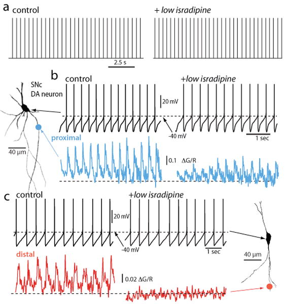
a. Digitized cell-attached patch recordings from an SNc DA neuron before and after application of isradipine (5 μM). The median discharge rate before isradipine application was 2.2 Hz and 2.4 Hz after (p>0.05, n=4). b. Whole cell recording from the cell shown to the left (projection image) before and after isradipine (5 μM) application; there was no significant change in discharge rate in this cell or in 10 others. At the bottom, 2PLSM measurements of Fluo-4 fluorescence (G) at a proximal dendritic location (∼40 μm from the soma) normalized by the fluorescence of the red Alexa dye used to image the cell. c. Somatic recording during imaging at a more distal dendritic location (∼120-200 μm from the soma). Note the complete elimination of the spike associated dendritic Ca2+ transient at the distal imaging site. Similar results were obtained in 6 other neurons. From [46]