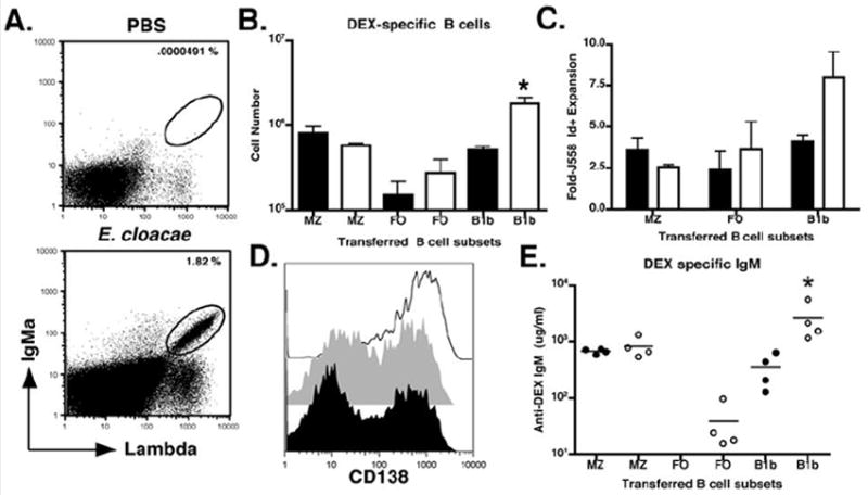
Figure 8. Role of splenic MZ, FO, and peritoneal, B1b B cells in DEX specific antibody response.

(A) Representative FACS profile of B6 mice reconstituted with either splenic MZ, FO, or peritoneal B1b B cells from naïve or E. cloacae primed VHJ558 TG mice. Mice were immunized with E. cloacae 24 hours after reconstitution with each B cell population. (B) Numbers of IgMa+ λ1+ B cells recovered from the spleens of B6 recipient mice reconstituted with naïve MZ (91 +/- 13% TD6-4+), FO (25 +/- 10% TD6-4+), B1b (50 +/- 15% TD6-4+) (black) or antigen experienced (white) MZ (90 +/- 15 TD6-4+), FO (30 +/- 10% TD6-4+), or B1b B cells (90 +/- 10% TD6-4+) 3 days after E. cloacae immunization. (C) To calculate the fold expansion, numbers of J558 Id+ B cells in recipient mice 3 days after E. cloacae immunization. Numbers of J558 Id+ B cells derived from naïve (black) and antigen experienced (white) MZ, FO, and B1b reconstituted recipients immunized with E. cloacae were calculated and divided by the initial numbers of the corresponding populations (D) CD138 expression by IgMa+ λ1+ B cells recovered from B6 recipient mice reconstituted with naïve or antigen experienced MZ (black line), FO (grey histogram), and B1b (black histogram) B cell subsets 3 days post E. cloacae immunization. (E) DEX specific IgMa antibody titers in recipient mice at 3 days post E. cloacae immunization for primary (closed circles) and secondary (open circles) DEX-specific antibody production. The amount of DEX-specific antibodies in the serum were displayed as micrograms/milliliter or ug/ml. All FACS plots and data (mean +/- SEM) are representative of 3 independent experiments containing 3 mice per group.
