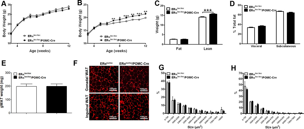
Figure 4.
Deletion of ERα in POMC neurons leads to increased body weight and lean mass. (A) Weekly body weight in male mice weaned on regular chow (n=33 or 34/genotype). (B) Weekly body weight in female mice weaned on regular chow (n=25 or 32/genotype). (C) Body composition in 11-week old female mice fed with regular chow (n=25 or 32/genotype). (D) Relative fat distribution in the visceral and subcutaneous depots in 18-week old female mice fed with regular chow (n=5/genotype). (E) Weight of gonadal WAT in 6-week old female mice fed with regular chow (n=8 or 9/genotype). (F) Representative photomicrographs of H&E staining of gonadal WAT and inguinal WAT from 5-month old chow-fed females. (G–H) Cell size in gonadal WAT (G) and inguinal WAT (h) from 5-month old chow-fed females (n=3/genotype). Data are presented as mean ± SEM, and * P<0.05, **P<0.01 and ***P<0.001 between ERαlox/lox/POMC-Cre mice and ERαlox/lox mice.

