
Figure 2.
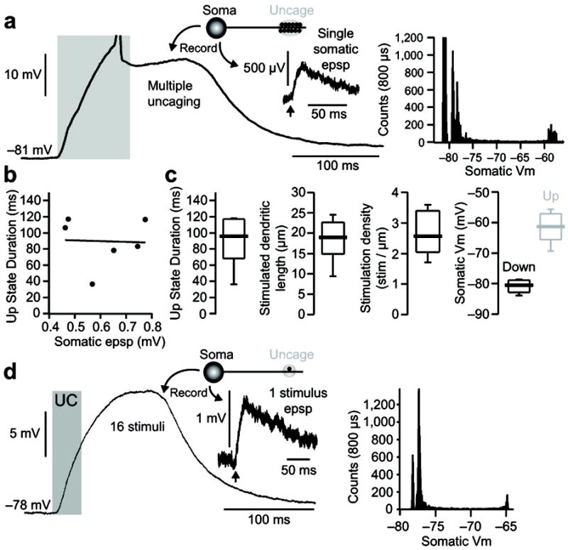
State transitions generated by proximal and distal dendritic glutamate uncaging are independent of the somatic EPSP size and recording internal used. (A) Example of a state transition induced in a dSPN: recording was made using a K-gluconate based internal: (in mM) 115 K-gluconate, 20 KCl, 10 Na-phosphocreatine, 10 HEPES, 2 Mg-ATP, and 0.3 Na-GTP)47 and Fluo 4FF; laser power was tuned to evoke small (463 μV) somatic EPSPs from a 0.5ms stimulation of a single distal dendritic spine (inset). The corresponding all points histogram for this cell is shown to the right. (B) Once a state transition is achieved, the up-state duration is not correlated with either the somatic EPSP induced by stimulation of a single distal dendritic spine or the recording internal solution used (n=6 cells; internal = KMeSo4 or K-gluconate with Fluo 4 or Fluo 4FF). (C) Though requiring a higher stimulation density, up-states induced using small amplitude (463–774 μV; n=6 cells) somatic EPSP are similar to those induced by supra-mV EPSP (i.e. figure 1). (D) In one case a modest state transition was produced by the repetitive stimulation of a single distal spine (16 stimuli, 0.5 ms duration, 500 Hz). Average somatic EPSP evoked by a single stimulus and the corresponding all points histogram are shown to the right.