
Figure 4.
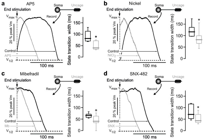
State transition generation requires NMDA receptors and voltage gated Ca2+ channels. (A) State transitions were generated in dSPNs by glutamate uncaging on distal dendritic spines, and then the response to the same uncaging protocol was measured in the presence of the NMDA receptor antagonist AP5 (100 μM). Shaded region indicates timing of glutamate uncaging. Voltage traces are normalized to maximum amplitude and overlain. Traces shown to the right are aligned to the end of the last uncaging pulse and normalized to the maximum amplitude of the state transition, as shown in figure 1d. Box plots indicate time between end of uncaging stimulation and 25% voltage fall. AP5 significantly reduced state transition duration, abolishing it in most cases (n=5, p<0.05 Mann-Whitney rank sum test). (B–D) State transitions were generated in dSPNs by glutamate uncaging on distal dendritic spines, and then the response to the same uncaging protocol was measured in the presence of (B) NiCl2 (50 μM; n=8), (C) Mibefradil (20 μM; n=8) or (D) SNX-482 (0.3 μM; n=6). Box plots indicate time between end of uncaging stimulation and 25% voltage fall. All three agents significantly reduce the somatic state transition duration (* p<0.05 Mann-Whitney rank sum test).