
Figure 5.
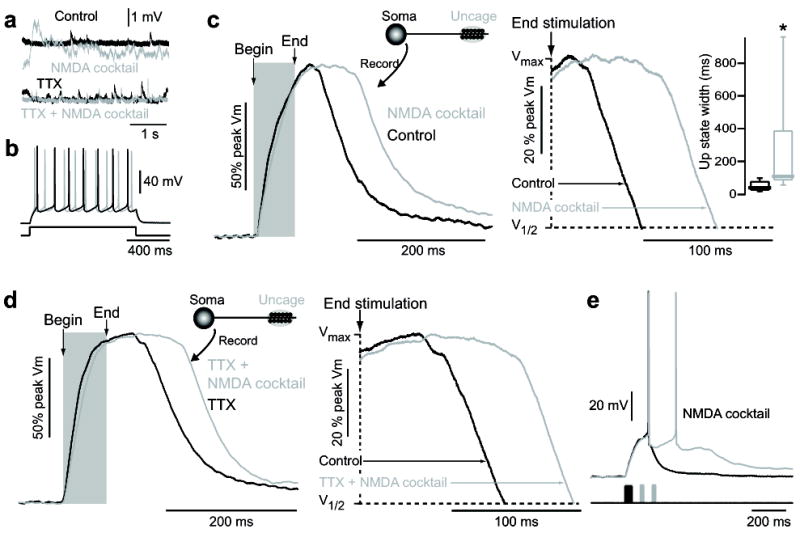
NMDA acts at distal dendritic synapses to enable state transition generation. (A) Somatic voltage recordings of spontaneous depolarizations in a dSPN before and after application of 10 μM NMDA + 10 μM glycine + 10 μM gabazine (SR95531) (“NMDA cocktail”, top) shows an increase in excitatory inputs, which is prevented by 1 μM TTX (bottom). (B) Somatic voltage of a dSPN in response to suprathreshold current injection in the presence and absence of 10 μM NMDA/glycine/gabazine. No regenerative changes in somatic voltage were observed, implying both the location of up-state duration and the NMDA receptors involved in their maintenance are located distally. (C) State transitions were evoked by glutamate uncaging on distal dendritic spines of dSPNs, then again in the presence of 10 μM NMDA/glycine/gabazine. Somatic up-state duration was significantly enhanced (n=6, p<0.05 Signed Rank test). Shaded region indicates timing of glutamate uncaging. Traces are normalized to the maximum amplitude. Box plots indicate time between end of uncaging and 25% voltage fall. (D) The increase in up-state duration in dSPNs caused by 10 μM NMDA/glycine/gabazine is not occluded by 1 μM TTX, suggesting the action of NMDA is directly on distal SPN dendrites and does not rely upon action potential dependent glutamate release (n=4). (E) A state transition was induced by stimulation of 10 distal dendritic spines (twice each) in a dSPN. The up-state could be maintained by supplemental stimulation of these same spines (once each) every 50 ms. Uncaging pulses are indicated by shaded regions.