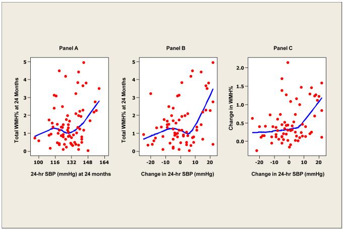
Figure 1. White matter hyperintensity and ambulatory blood pressure.
Locally weighted scatterplot smoother plots of 24-hour average systolic BP and white matter hyperintensity (WMH) lesions (as percent of total intracranial volume) (panel A), change in 24-hour systolic BP and WMH (%) (panel B) and change in 24-hour systolic BP and change in WMH (%) at 24 months (panel C).

