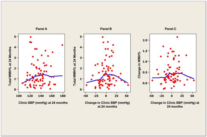
Figure 2. White matter hyperintensity and clinic blood pressure.
Locally weighted scatterplot smoother plots of clinic systolic BP and white matter hyperintensity (WMH) lesions (as percent of total intracranial volume) (panel A), change in clinic systolic BP and WMH (%) (panel B) and change in clinic systolic BP and change in WMH (%) at 24 months (panel C).

