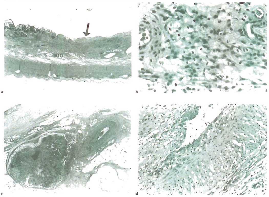
Fig. 5.
a) CR of human small bowel allograft shows focal, non-healing ulcers(arrow), as shown here, and thickening and fibrosis of the submucosa(sm). b) A higher magnification of the submucosa reveals abundant foam cell deposition, similar to that seen in the arterial intima of blood vessels with OA. c) Like other allografts, the arteries affected by OA are not commonly sampled in biopsies. This section shows a mesenteric artery with OA near a mesenteric lymph node with lymphold depletion and fibrosis(arrow). d) A higher magnification shows mild lymphocytic intimal inflammation, intimal foam cell deposition and endothelial cell hypertrophy. Note also the marked medial vacuolization, which is due to foam cell infiltration between myocytes and intercellular edema.

