Fig. 6.
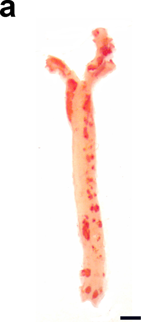
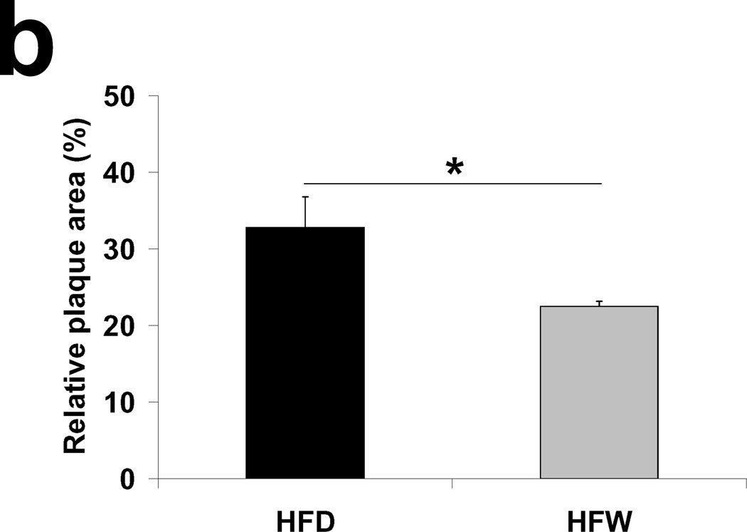
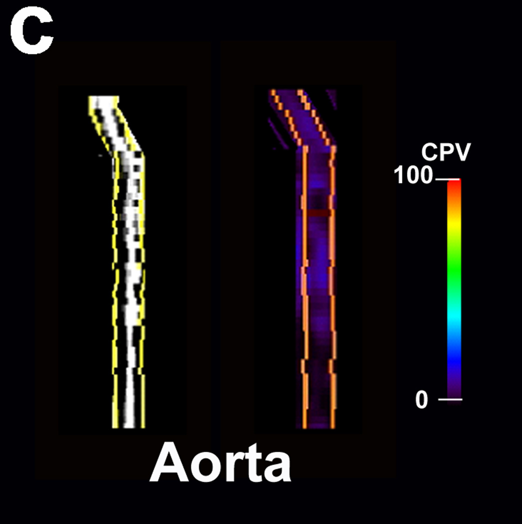
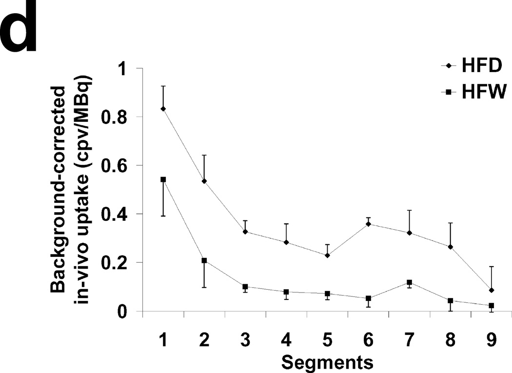
A) Representative example of Oil Red O staining of the aorta of an apoE−/− mouse fed high fat diet for 2 months followed by normal chow for one month. Scale bar: 2mm. B) Relative plaque area detected by Oil Red O staining of aortae of animals fed high fat diet for 3 months (HFD) or high fat diet for 2 months followed by 1 month of normal chow (HFW). n=3 in each group. *: p=0.01. C) Representative examples of multiplanar reconstruction of SPECT (left) and CT (right) images of aorta in the HFW group. D) MicroSPECT-derived quantification of RP782 uptake in HFD and HFW animals. n=4 in each group. cpv: counts per voxel.


