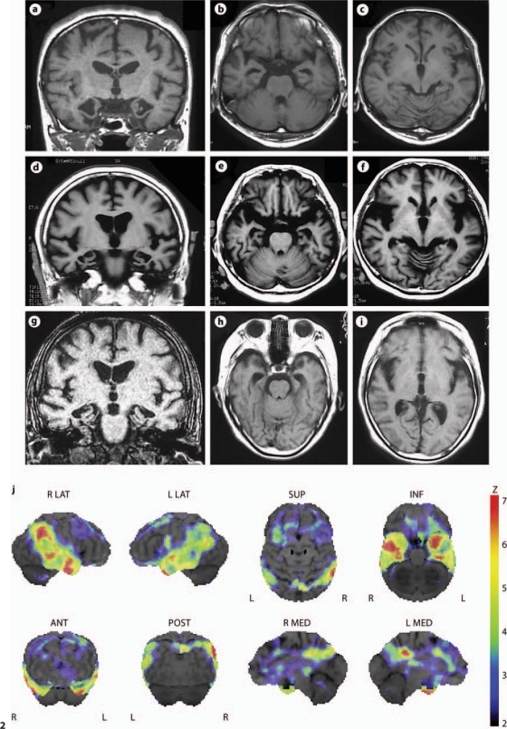
Fig. 2.
Representative T1-weighted MR images of 3 patients: Ped 3367-P1 at the age of 56 years (a–c); Ped 3367-P3 at the age of 67 years (d–f), and Ped 4550-P1 at the age of 77 years (g–i). Coronal images revealed marked atrophy of the medial temporal lobe including the hippocampus and parahypocampal gyrus (a, d, e). On axial images, an enlarged inferior horn of the lateral ventricles was observed (b, e, h). Atrophic change in the frontal lobe is less noticeable (c, f, i). j Decreased rCBF by 123I-IMP SPECT with 3D-SSP in Ped 3367-P3 at the age of 67 years. After global normalization to the mean blood flow for the entire brain, rCBF in the patient was compared with that in normal controls by the Z test. Color-coding represents the statistical significance (Z score) of the decrease in rCBF. Decreases in rCBF were observed in the bilateral temporal lobes as well as in the cingulate gyrus of anterior (ANT) and posterior (POST) portions. L = Left; R = right; INF = inferior; LAT = lateral; MED = medial; SUP = superior.

