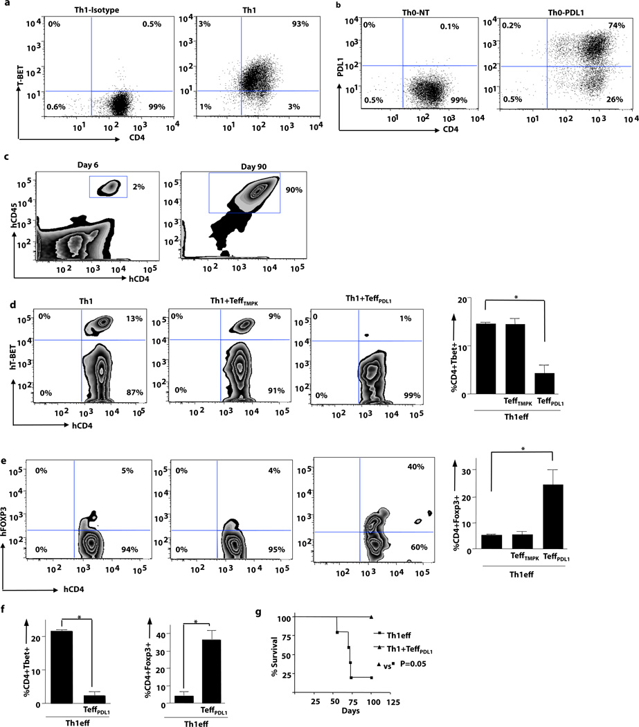
Figure 3. Th1 Cells Mediate Lethal xGVHD: Abrogation by PDL1- expressing Effector T Cells.
(a) Human Th1 cells were expanded ex vivo. Prior to adoptive transfer, Th1 cells were evaluated by intracellular flow cytometry for transcription factor expression and cytokine content (b) Prior to adoptive transfer, transduced human T cells were evaluated for PDL1 expression (non-transduced cells; PDL1-transduced cells). (c) NSG mice were injected with Th1 cells (1 × 10⁶) alone or with PDL1-transduced T cells (5 × 10⁴). Human T cell splenic engraftment for recipients of Th1 cells plus PDL1-transduced cells is shown at day 6 and day 90 after transfer. (d) TBET expression by intracellular flow cytometry was monitored in cohorts that received Th1 cells alone, Th1 cells plus control LV-transduced T cells, or Th1 cells plus PDL1-LV transduced T cells. Right panel shows pooled data for TBET expression for each cohort. (e) FOXP3 expression by i.c. flow cytometry was monitored at day 6 after transplant in cohorts that received Th1 cells alone, Th1 cells plus control LV-transduced T cells, or Th1 cells plus PDL1-LV transduced T cells. Right panel shows pooled data for FOXP3 expression for each cohort. (f) TBET and FOXP3 expression at day 90 after transplant in recipients of Th1 cells or Th1 cells plus PDL1-expressing T cells. (g) Survival curve for recipients of Th1 cells alone or Th1 cells plus PDL1-expressing T cells. (* indicates p ≤ 0.05, ** indicates p ≤ 0.005).

