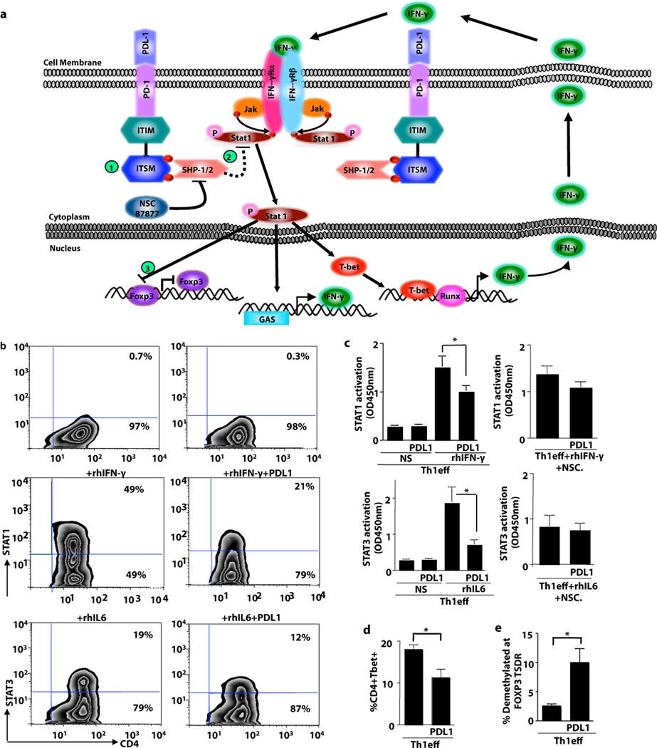
Figure 6. PDL1 Induction of Th1 Cell Plasticity Associates with STAT1 Inactivation.
(a) Working model for Th1 cell plasticity: (1) PDL1/PD1 interactions recruit SHP1/2; (2) SHP1/2 inactivates STAT1, thereby abrogating IFN-mediated maintenance of TBET; (3) STAT1 inhibits FOXP3. (b) To investigate aspects of this model, Th1 cells were restimulated in the presence of IFN-γ or IL-6 for 30 min prior to phosphoflow cytometry. Representative examples shown are Th1 cells without additional cytokine addition either alone or with PDL1-coated beads; Th1 cells plus a STAT1 activating cytokine (IFN-γ) alone or with PDL1-coated beads; and Th1 cells plus a STAT3 activating cytokine (IL-6) alone or with PDL1-coated beads. (c) Nuclear lysates of Th1 cells were obtained (n=5 donors) and subjected to an ELISA-based STAT activation assay. STAT1 activation was evaluated in the presence of PDL1 (top left panel) or PDL1 plus the SHP1/2 inhibitor NSC87877 (top right panel); STAT3 activation was evaluated in the presence of PDL1 (bottom left panel) or PDL1 plus NSC87877 (bottom right panel). (d) Th1 cells were evaluated for TBET expression in the presence or absence of PDL1. (e) Th1 cells were evaluated for demethylation status of the FOXP3 TSDR locus. (For each panel: * indicates p ≤ 0.05, ** indicates p ≤ 0.005, *** indicates p ≤ 0.0005).

