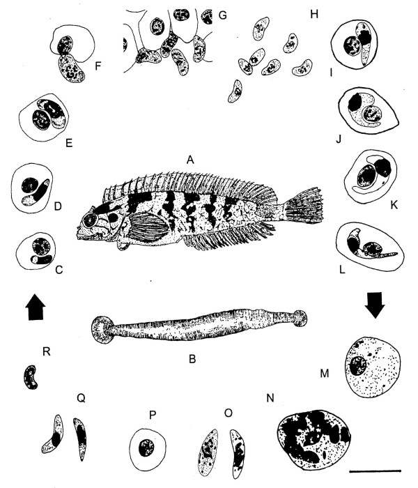
Figure 1.
A-R. Life cycle of Haemogregarina curvata. A: Clinus cottoides redrawn from Penrith [26] (not drawn to scale). B: Adult Zeylanicobdella arugamensis redrawn from De Silva [27] (not drawn to scale). C-R: Microscope drawings to illustrate the life cycle of Haemogregarina curvata. C-L: Stages within peripheral blood smears from C. cottoides. M-R: Stages within Zeylanicobdella arugamensis squashes and histological sections through salivary glands. Arrows indicate stages of suspected transfer of haemogregarine from one host to another. C: Small intraerythrocytic trophozoite. D: Larger trophozoite. E: Intraerythrocytic meront. F: Extracellular meront. G, H: Extracellular merozoites. I: Intraerythrocytic pregamont form. J: Immature gamont. K Intermediate gamont. L: Mature intraerythrocytic gamont. M Immature oocyst. N Developing oocyst with 8-10 nuclei. O: Free sporozoites. P: Meront. Q: First generation merozoites. R: Second generation merozoite. Scale bar = 10 μm.

