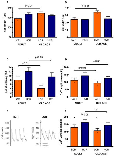
Figure 2. Properties of cardiac ventricular cardiomyocytes were more compromised as a function of age in LCR compared with HCR rats.
(A&B) Cell length and width increased in old LCR but not in old HCR. (C) Isolated cell shortening greater in HCR than LCR in both adult and old age (D) Amplitude of Ca2+ transients decreased with aging in LCR but not HCR. (E) Example signals of Ca2+ transients. (F) Sarcoplasmic reticulum Ca2+ load measured after caffeine application was reduced in LCR vs. HCR cells, and deteriorated with aging in LCR, but not HCR cells. Adult: 15-20 months; Old Age: >25 months. *: p<0.05 age-matched LCR vs. HCR.

