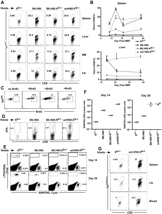
Figure 1.
LTFNYRNL derived from H60 is efficiently cross-presented. (A-D) Kb−/−, B6.H60, B6.H60.Kb−/−, and actH60 mice were irradiated and reconstituted with 6 × 10⁶ BM cells, 2.4 × 10⁶ CD8 cells, and 105 CD4 cells from C3H.SW (Ly9.1+) mice. (A) Representative flow cytometry at day 14 after BMT. (B) Percentage of Ly9.1+CD8+ cells in spleen (top panel) and liver (botom panel) that were TetH60+ (3 mice per group at each time point); bars represent SE. (C) Representative BrDU staining of Ly9.1+CD8+TetH60+ cells in actH60.Kb−/− recipients after a single BrDU pulse 2 hours before death at day 14. Each plot is from an individual mouse. (D) Representative staining for intracellular IFN-γ in LTFNYRNL-stimulated splenocytes harvested at day 14 (gated on live Ly9.1+CD8+ cells). (E-F) Mice were transplanted as in panels A through D; and on days 14 and 28, recipients were injected with LTFNYRNL- or SIINFEKL-pulsed C3H.SW splenocytes labeled with CFSE or cyan, respectively. Eighteen hours later, splenocytes were analyzed for the presence of CFSE+ and cyan+ cells. Representative flow cytometry at day 14 (E) and the ratios of cyan+/CFSE+ cells (F; each symbol is from an individual mouse). CD8 cells in actOVA.Kb−/− mice transplanted as in panels A to D mount a strong response to SIINFEKL (G). Shown is representative flow cytometry of spleen, LN, and blood cells at day 14 (gated on donor CD8+ cells). Data are representative of at least 2 experiments with similar results.

