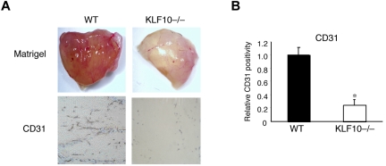
Figure 6.
Reduced angiogenesis in Matrigel plugs implanted in KLF10−/− mice. (A-B) Matrigel plugs subcutaneously implanted for 8 days in WT or KLF10−/− mice (n = 10 per group) were stained for CD31. (A) Angiogenesis in whole Matrigel plugs. Matrigel images were photographed with an Olympus, Model SZ61 camera (top). CD31 staining in paraffin sections (5 μm; bottom) was analyzed with an Olympus, Fluoview, Model FV1000 camera at 10× magnification and FV10-ASW Version 02.01 software and quantitated as relative CD31 positivity (B). *P < .01 versus WT.

