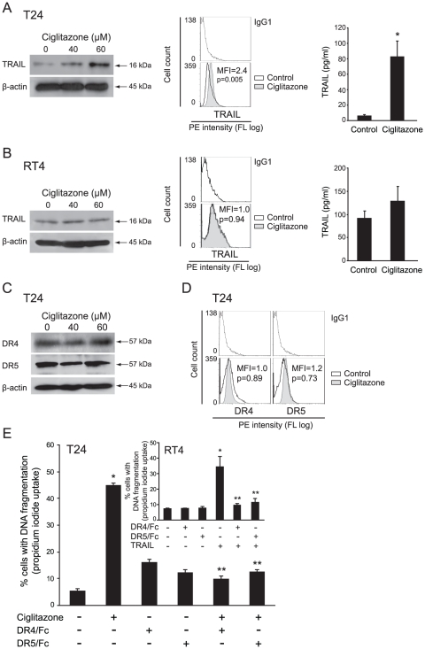
Figure 4. Effect of ciglitazone on soluble and membrane-bound TRAIL and on DR4/DR5 receptors signaling pathway.
RT4 and T24 cells were treated for 24 h with ciglitazone at 40 and/or 60 µM. In RT4 (A) and T24 (B) cells, whole cell lysates were assayed for TRAIL expression by western-blotting analysis. β-actin was used as an internal loading control. Cells were stained with anti-TRAIL-PE and analysed by flow cytometry. After 40 µM ciglitazone treatment for 24 h, conditioned media were collected and the concentration of TRAIL was measured by ELISA. Data are means ± SD of 2 independent experiments performed in quadruplicates. *P<0.05, significant differences compared with untreated cells with the use of two-tailed unpaired Student's t test. (C) Cellular proteins were isolated from T24 cells and subjected to immunoblotting for detection of DR4 and DR5. β-actin was used as an internal loading control. (D) T24 cells were exposed to 40 µM ciglitazone for 12 h, stained with anti-DR4-PE or anti-DR5-PE and analysed by flow cytometry. (E) T24 cells were preincubated with or without monoclonal antibodies blocking DR4 and DR5 receptors (5 mg/ml) for 1 h and stimulated for 12 h by 40 µM ciglitazone or 50 ng/ml TRAIL for RT4 cells (insert). The percentage of cells showing hypodiploid DNA content (sub-G1 peak) was evaluated by flow cytometry analysis. Data are means ± SD of 3 independent experiments performed in triplicates. *P<0.05, significant differences compared with untreated cells ; **P<0.05, significant differences compared with ciglitazone-treated cells with the use of two-tailed unpaired Student's t test.

