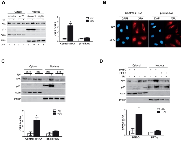
Figure 1. p53 is required for the XPA nuclear import upon UV irradiation.
A, p53 was transiently knocked down with siRNA duplexes in HeLa cells. After treatment with or without 20 J/m2 UV followed by a 2-hr recovery, subcellular fractionation and Western blotting were performed to assess the re-distribution of XPA. β-actin and PARP were probed as cytoplasmic and nuclear protein controls, respectively. The quantitative data were obtained from at least three independent experiments. nXPA/cXPA represents the ratio of nuclear XPA to cytoplasmic XPA. B, Immunofluorescence microscopic analysis of cells transfected with control or p53 siRNA and with or without UV irradiation. C, A549/LXSN(p53+) and A549/E6(p53−) cells were mock- or UV-irradiated. Cytosol and nuclear fractions were collected and analyzed by Western blotting. D, A459 cells were pre-treated with pifithrin-α (30 uM), an inhibitor of p53 transcriptional activity, for 20 hrs. After UV irradiation and a 2-hr recovery, the cells were analyzed for subcellular localization of XPA. The * in the plots indicates a statistically significant (p<0.05) difference between the groups being compared.

