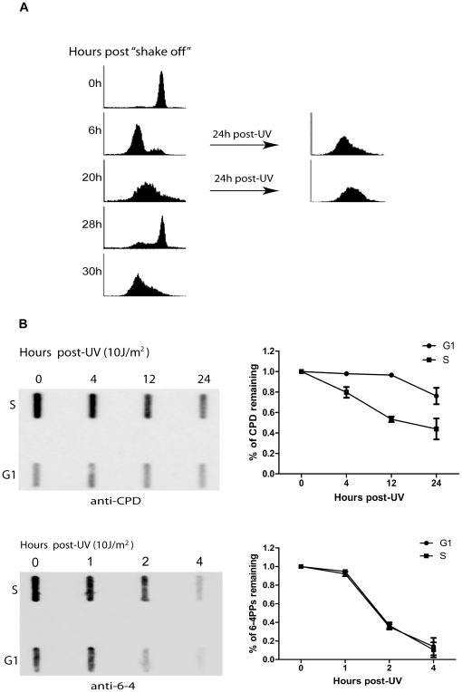
Figure 4. Removal of UV-induced DNA damage in G1- and S-phase cells.
A. Mitotically-synchronized HeLa cells were fixed and stained with propidium iodide at the indicated time points following the mitotic “shake off”. The cell cycle distribution then was analyzed by flow cytometry. Cells at G1 (at the 6 hours post-“shake off”) or S (20 hours post-“shake off”) phase, were UV irradiated at 10 J/m2, followed by a recovery of 24 hours. B. Cells at G1 or S phase were UV irradiated at 10 J/m2, followed by the indicated periods of repair. Cellular DNA were isolated and the removal of CPDs and 6-4PPs was measured by slot-blot assay. The amounts of CPDs or 6-4PPs were normalized to the values at zero hour and quantified based on three independent measurements.

