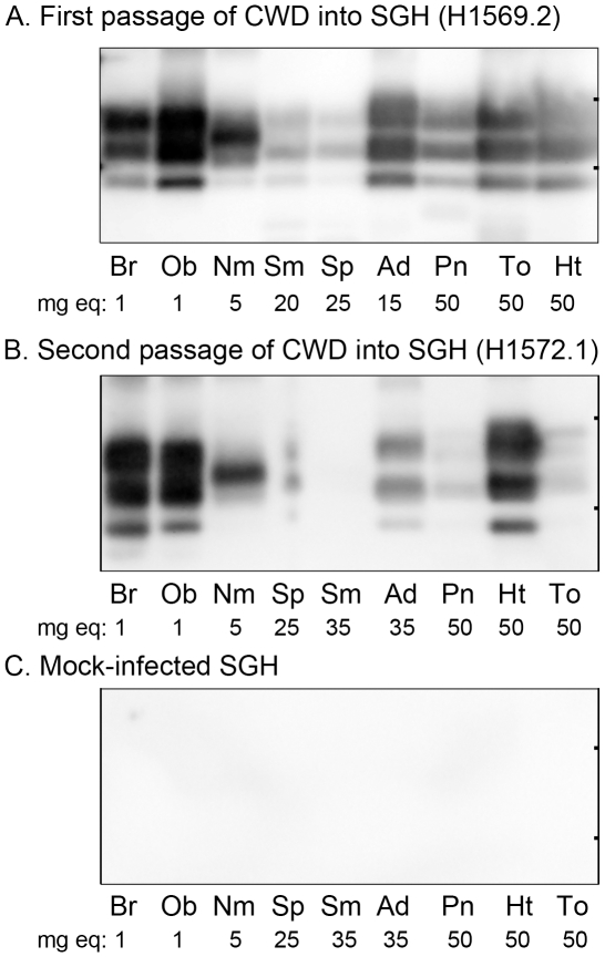
Figure 4. Western blot for PrPSc in central and peripheral tissues of Syrian golden hamsters infected with chronic wasting disease.
Tissues from hamsters infected with WST CWD after first (A) and second (B) passage (see Table 1, recipient groups H1569 and H1572) and mock infected brain (C) were enriched for PrPSc by differential ultracentrifugation and proteinase K digestion followed by NuPAGE and anti-PrP western blot analysis. The amount of tissue equivalents in milligrams (mg eq) loaded into each lane is indicated below each tissue. Key: Br, brain; Ob, olfactory bulb; Nm, nasal mucosa; Sm, submandibular lymph node; Sp, spleen; Ad, adrenal gland; Pn, pancreas; To, tongue; and Ht, heart. Tick marks on the right vertical frame of each panel indicate the 20 and 30 kDa molecular weight markers.

