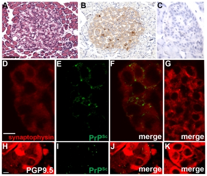
Figure 6. Immunohistochemistry for PrPSc in neuroendocrine tissues in Syrian golden hamsters infected with chronic wasting disease.
Pancreas (A through G) and adrenal gland (H through K) from mock (C, G, K) and WST CWD-infected (A, B, D, E, F, H, I, J) hamsters. Panels D through F, and H through J are the same field of view, which are separated into three panels according to the immunofluorescence staining. Pancreas was analyzed by dual immunofluorescence for synaptophysin (D, F, G) and PrPSc (E, F, G) while the adrenal gland was analyzed by dual immunofluorescence for PGP9.5 (H, J, K) and PrPSc (I, J, K) using laser scanning confocal microscopy. Serial sections of pancreas were also analyzed by hematoxylin and eosin (A) and PrPSc immunohistochemistry (B, C; brown punctate aggregates) and counterstained with hematoxylin. Scale bar in D and H is 10 µm and in A the scale bar is 100 µm.

