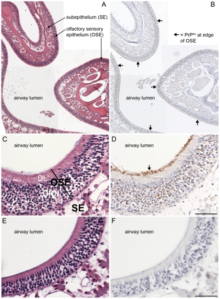
Figure 7. Distribution of PrPSc in nasal cavity in Syrian golden hamsters infected with chronic wasting disease.
Low (A, B) and high (C, D, E, F) magnification photomicrographs of the nasal cavity from Syrian golden hamsters infected with WST CWD brain (A, B, C, D) and mock-infected brain (E, F) were stained with hematoxylin and eosin (A, C, E,) or immunostained for PrPSc (brown deposit) and counterstained with hematoxylin (B, D, F). Key: OSE, olfactory sensory epithelium; SE, subepithelial layer; ORN, olfactory receptor neurons; and DL, dendrite layer. Arrows indicate PrPSc deposition at the border of the OSE layer and lumen of the nasal airway. Scale bar in D and F is 50 µm.

