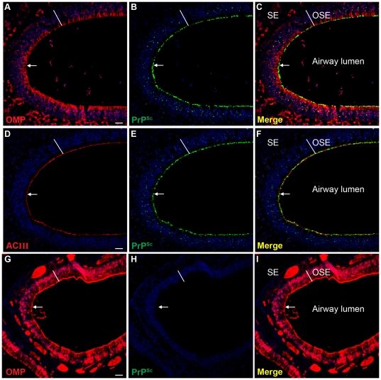
Figure 8. Distribution of PrPSc in olfactory sensory epithelium in Syrian golden hamsters infected with chronic wasting disease.
Laser scanning confocal microscopy of olfactory marker protein (OMP)(A, G), PrPSc (B, H), and for both OMP and PrPSc (Merge)(C, I) in Syrian golden hamsters infected with TgMo-sghPrP CWD (A, B, C) and mock-infected hamsters (G, H, I). Laser scanning confocal microscopy of adenylyl cyclase III (ACIII)(D), PrPSc (E), and for both ACIII and PrPSc (Merge)(F) in CWD infected SGH. Panels A through C, D through F and G through I are the same field of view that are separated into three panels according to the immunofluorescence staining. ToPro®-3 staining of nuclei is indicated by blue fluorescence. Olfactory receptor neurons in the olfactory sensory epithelium (OSE width indicated by white line), and nerve fibers in the subepithelial layer (SE), both express high levels of OMP (A, C and G and I). ACIII is located on the sensory cilia that project from the terminal dendrites of ORNs and its distribution was prominent at the border between the OSE and airway lumen (D and F). The white arrow points to the distal edge of the OSE where it borders the lumen of the nasal airway. Scale bar in A, D and G is 50 µm.

