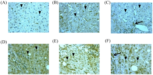
Figure 1. Immunohistochemistry for STAT1 in pretreated liver biopsy samples.
(A)–(C) Patients with achievement of SVR. (D)–(F) Patients without achievement of SVR. (A) From a patient infected with HCV G1, high viral load (SVR, IL28B rs8099917TT). Arrowheads show markedly reduced nuclear staining for STAT1. (B) From a patient infected with HCV G1, high viral load (SVR, IL28B rs8099917TT). Arrowheads show markedly reduced nuclear staining for STAT1. (C) From a patient infected with HCV G1, high viral load (SVR, IL28B rs8099917TG). Arrow, bile duct; arrowheads show markedly reduced nuclear staining for STAT1. (D) From a patient infected with HCV G1, high viral load (Null responder, IL28B rs8099917TG). Arrowheads show more distinct nuclear staining for STAT1. (E) From a patient infected with HCV G1, high viral load (Relapser, IL28B rs8099917TT). Arrowheads show more distinct nuclear staining for STAT1. (F) From a patient infected with HCV G1, high viral load (Relapser, IL28B rs8099917TT). Arrow, bile duct; Arrowheads show more distinct nuclear staining for STAT1.

