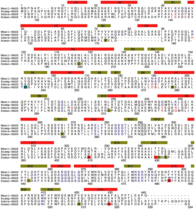
Figure 5. Sequence alignment of mmNAGS/K, xcNAGS/K, zebra fish NAGS and human NAGS.
The sequence encoding secondary structure elements are indicated by boxes in yellow-green (β-strand) and red (α-helix). Encoded amino acids that are modeled in binding of ligands (ATP, NAG, AcCoA, glutamate, L-arginine), are indicated in blue. The linker residue, Gly291, is boxed. The missense human mutations are indicated by boxes in red, yellow-green, light blue for neonatal, late-onset and unknown onset patients, respectively.

