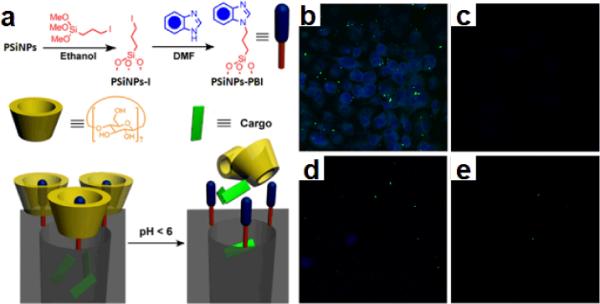
Fig. 11.

(a) Schematic illustration of pH-operated mechanism of the nanovalves. Confocal images of PANC-1 cells incubated with porous silicon nanorods: (b) Cells treated with 20 μg/ml of FITC-PSiNPs loaded with Hoechst 33342 at 37 °C. (c) Same experiment conducted at 4 °C No staining was observed after incubation at low temperature. (d) and (e) Competition tests: cells were treated with 20 μg/ml of FITC-PSiNPs loaded with Hoechst 33342 and (d) 60μg/ml and (e) 200μg/ml of plain PSiNPs at 37 °C. (Adapted from ref. 51. Copyright 2011 American Chemical Society.)
