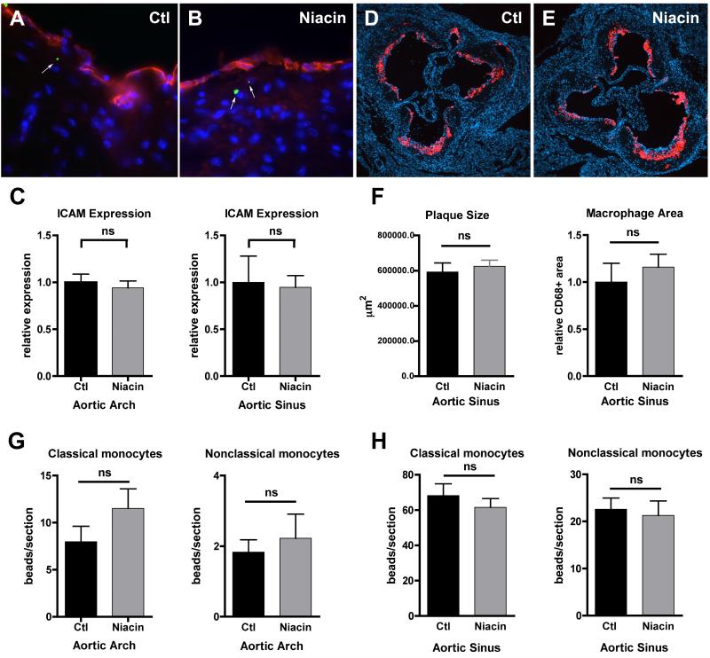
Figure 1.
Effect of niacin treatment on monocyte entry into advanced atherosclerotic lesions. A cohort of apoE−/− mice was transitioned to a HFD supplemented with 1% niacin (Niacin) for two weeks while the other half remained on control HFD (Ctl). (A-B) Representative images of aortic sinuses from mice with green microbead-labeled nonclassical monocytes (green microspheres, white arrows) and stained with anti-ICAM antibody (red) and DAPI (blue), 63X magnification. (C) Graphs depict quantification of anti-ICAM antibody staining in the aortic arch and sinus. (D-E) Representative images of aortic sinuses stained with anti-CD68 antibody (red) to depict macrophages and DAPI (blue), 10X magnification. (F) Graphs depict quantification of plaque size and macrophage area in aortic sinuses. (G-H) Classical or nonclassical monocytes were labeled intravenously following niacin treatment. Mice were sacrificed 2-5 days post-labeling, aortic arches and hearts removed and sectioned and beads per section in the (G) aortic arch and (H) sinus enumerated. There were no statistically significant differences (ns), as determined by an unpaired T-test, in ICAM staining, morphometric measurements, or the number of beads in any treatment group (Ctl vs Niacin) in any experiments. N=4-9/group, each experiment repeated 2-4 times.

