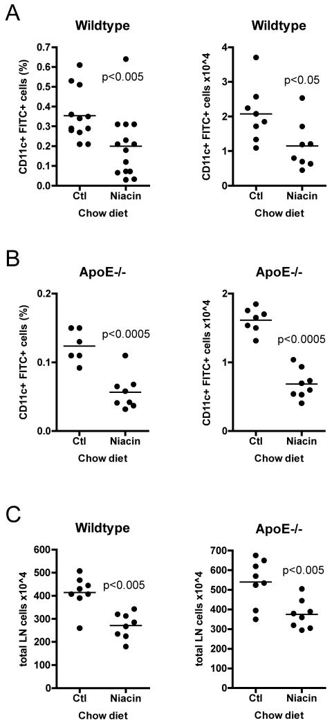
Figure 2.
Effect of niacin therapy on the migration of DCs from skin to draining lymph nodes. Mice were fed control chow (Ctl) or 1% niacin-supplemented chow (Niacin) for two weeks. 18 hours after FITC-containing contact sensitizer solution was applied to the shaved flanks of anesthetized mice, animals were sacrificed, brachial and axillary lymph nodes were removed and pooled by side, and FITC+ migrated DCs quantified by flow cytometry in (A) wild-type or (B) apoE−/− mice. (C) Total lymph node cellularity was quantified by manual counting. Each dot represents a pool of brachial and axillary lymph node from one side of a mouse (two dots per mouse). Each experimental condition was repeated 3-6 times with 3-5 mice per condition. P-values, depicted within each graph, were determined with an unpaired T-test.

