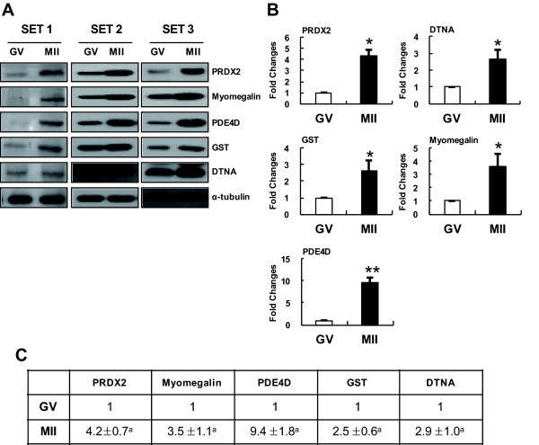
Figure 6.
Western-blot analyses confirming the upregulation of particular proteins during the M II stage. (A) Western blot analyses of total protein extracts from GV and M II oocytes (lane 1, GV oocytes; lane 2, M II oocytes) using specific antibodies against proteins (PRDX2, Myomegalin, PDF4D, GST and DTNA) identified by 2-DE. Alpha-tubulin was used as a loading control. (B) Quantification of PRDX2, Myomegalin, PDF4D, GST and DTNA expression in GV and MII embryos. PRDX2, Myomegalin, PDF4D, GST and DTNA protein expression increased following MII embryos. *Value significantly differs from the control (*P <0.05 and **P <0.01). The results, which were analyzed by the TINA program, are presented in a bar graph.

