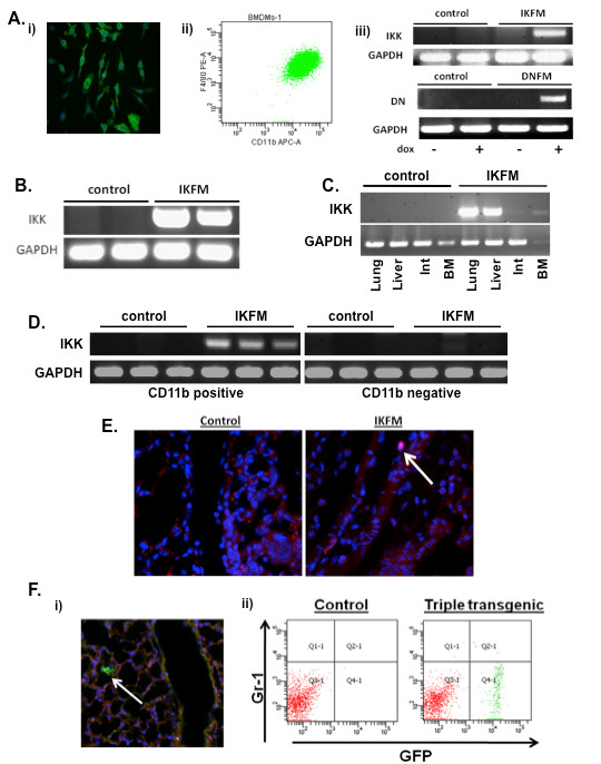
Figure 1.

Characterization of IKFM and DNFM mouse models. (a) Bone marrow-derived macrophages were isolated from IKFM and DNFM mice and (i) stained for F4/80 via immunofluorescent staining (green = F4/80 and blue = DAPI), (ii) subjected to flow cytometric analysis of F4/80 and CD11b and (iii) treated with 1 μg/ml doxycycline (dox) for 24 hours in vitro and gene expression analyzed via RT-PCR. (b) Peritoneal macrophages were isolated from IKFM mice treated with dox (2 g/L) for one week and transgene expression analyzed via RT-PCR. (c) IKFM mice and controls were treated with dox (2 g/L) for four weeks and transgene expression was detected by RT-PCR in lung, liver, intestines (Int), and bone marrow (BM). (d) IKFM and control mice were treated with dox (2 g/L) for one week and CD11b positive and negative populations isolated from lungs for RT-PCR analysis of IKK. GAPDH was performed as control. (e) Phospho-p65 (Ser536) immunofluorescent staining (red = phospho-p65 and blue = DAPI) of lungs from IKFM and control mice bearing lung metastases and treated with dox (2 g/L). (f) (i) Lung sections of TG/Cre/FMR mice treated with dox for one week (blue = DAPI) and (ii) flow analysis of TG/Cre/FMR and control lungs. Total cells were first sorted for CD45 and CD11b double positive cells. This double positive population was then further sorted for Gr-1 and GFP, as shown.
