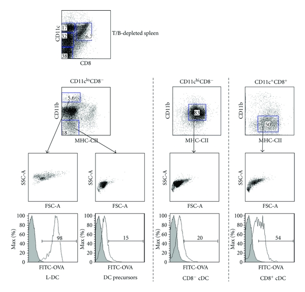
Figure 3.

Isolation of L-DC (LIVE-DC) in spleen amongst other DC subsets. Spleens were depleted of T and B cells using antibody-coated magnetic beads and then stained with antibodies specific for CD11c, CD11b, CD8α and MHC-II ahead of flow cytometric analysis. DC subsets were identified based on multiple parameters of forward scatter (FSC), side scatter (SSC), marker expression and endocytosis after intravenous administration of FITC-OVA (0.6 mg/mouse) 24 hours previously.
