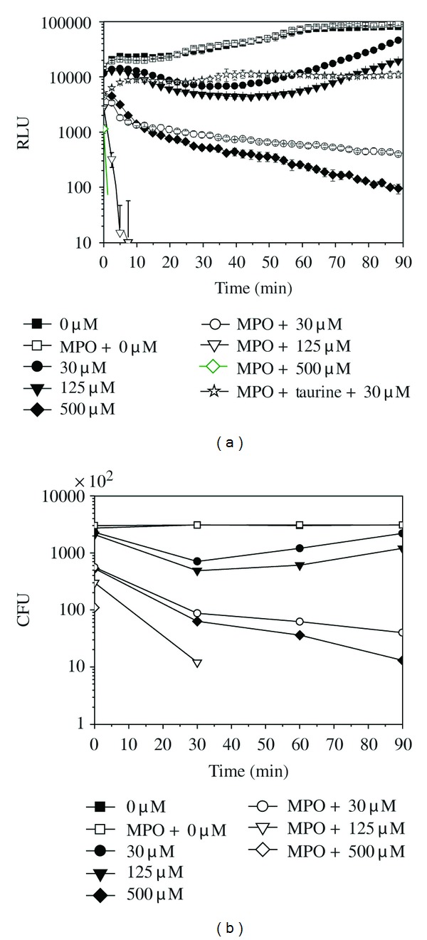
Figure 1.

The bioluminescence signal (a) and colony forming units (CFU)/200 μL (b) of E. coli-lux (3 × 10⁵ cells) incubated in the presence of various amounts of H2O2 (μM) and 1 μg/well MPO in phosphate buffer at 37°C. (■) 0 μM, (●) 30 μM, (▾) 125 μM, (♦) 500 μM, (□) MPO + 0 μM, (○) MPO + 30 μM, (∇) MPO + 125 μM, (♢) MPO + 500 μM, and (□) MPO + 50 mM of taurine + 30 μM. Relative luminescence unit (RLU) values are shown as the mean ± SD of measurements from three parallel wells.
