
Figure 1.
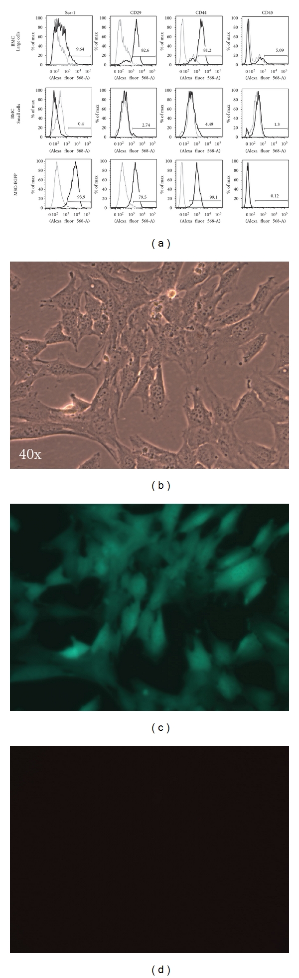
Phenotypic profiling of cultured BMCs and MSC-EGFPs. (a) BMCs cultured for three weeks in serum-containing mitogen-free conditions and mitogen-activated MSC-EGFPs at passage 18 were analyzed by Flow cytometry (FCM) using the Mouse Multipotent Mesenchymal Stromal Cell Marker Antibody Panel (R&D Systems). Shown are the positively staining markers Sca-1, CD29, and CD44, and an example of negative staining for the hematopoietic marker CD45. ((b), (c), (d)) Undifferentiated mesenchymal stromal cells derived from adult EGFP transgenic mice (MSC-EGFP) appear as large, flat fibroblast-like cells that express high levels of the enhanced green fluorescent protein reporter, EGFP. MSC-EGFPs are shown under (b) phase contrast and (c) GFP bandpass filter, demonstrating eGFP expression and (d) Texas Red bandpass filter, demonstrating lack of autofluorescence.